Comprehensive Eye and Vision Exam
Comprehensive Eye and Vision Exam
A regular eye examination is an essential preventive measure to protect vision, as many sight-threatening eye diseases have no obvious symptoms. Early diagnosis, timely referral, and appropriate treatment can help preserve vision.
Comprehensive eye exams can detect various eye diseases and disorders, such as glaucoma, cataracts, retinal detachments, and macular degeneration. They can also reveal systemic health conditions like diabetes and high blood pressure.
What a Comprehensive Eye Exam Includes:
1. History Taking
We begin by discussing your visual concerns, past eye examinations, treatments received, and any relevant medical, drug, and family history. We also analyze your visual needs at home, work, school, and during recreational activities to accurately determine your visual demands.
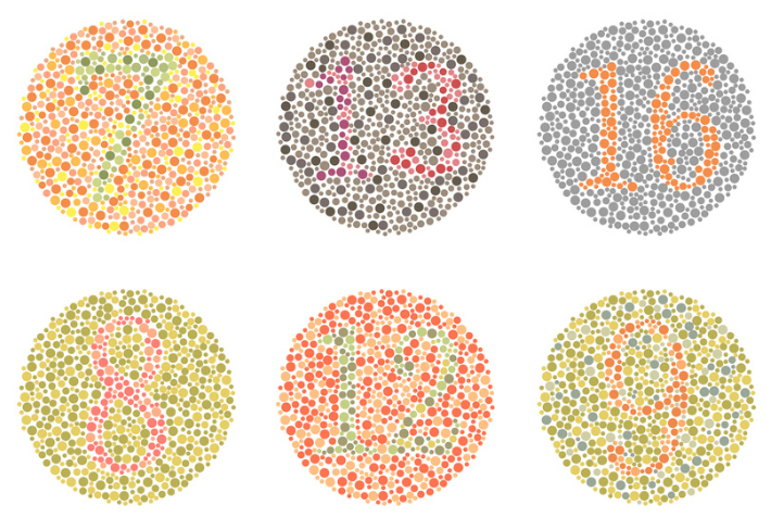
2. Preliminary tests
These tests assess:
- Colour vision
- Depth perception
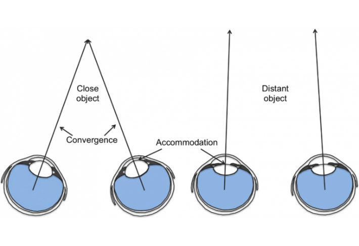
- Eye muscle movement
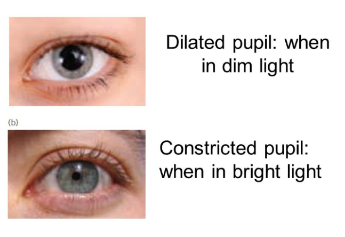
- Pupil response
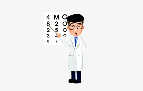
- Visual acuity
These evaluations provide a baseline for how well your eyes function and coordinate. Further testing will focus on any detected weaknesses.
3. Objective and Subjective Refraction
Refraction determines the lens power needed to correct nearsightedness, farsightedness, astigmatism, and presbyopia.
- Auto-refraction: A quick, initial assessment of your refractive status.
- Subjective refraction: Lenses are placed in front of your eyes, and adjustments are made based on your responses to refine the prescription.
- Retinoscopy: Used for younger children or patients who cannot respond accurately, allowing us to estimate the required eyeglass prescription by analyzing how light reflects from the eye.
4. Eye Health Evaluation
The eye is divided into:
- Anterior segment (front 1/3): Includes the cornea, iris, ciliary body, and crystalline lens.
- Posterior segment (back 2/3): Includes the vitreous humor, retina, choroid, and optic nerve.
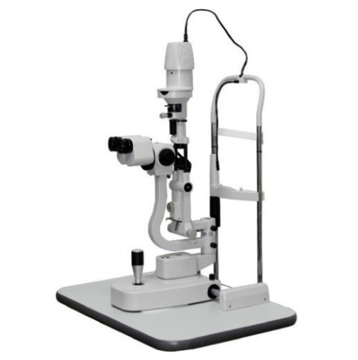
A) Slit Lamp Examination
This test assesses the anterior segment, allowing us to detect conditions like dry eyes, corneal opacity, and cataracts. It is also used to examine external structures such as the eyelids and eyelashes.
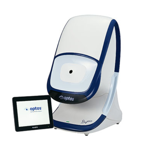
B) Fundus Photography (Optos Daytona / Clarus 500)
This imaging technology captures a 200° ultra-widefield view of the retina in a single scan. It helps detect retinal conditions such as:
- Age-related macular degeneration (AMD)
- Retinal degeneration (holes, breaks, or tears)
- Diabetic retinopathy
- Retinal vein/artery occlusion
- Posterior vitreous detachment
- Floaters
- Optic nerve atrophy
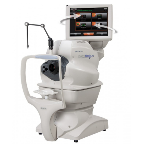
C) Optical Coherence Tomography (OCT)
OCT uses light waves to produce high-resolution images of the retina’s structural layers. It helps diagnose:
- Macular hole
- Macular edema
- Epiretinal membrane
- Central serous retinopathy
- Optic nerve abnormalities (glaucoma, optic nerve inflammation, or tumors)
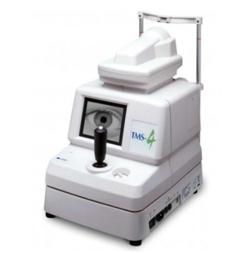
Additionally, corneal topography is used to detect keratoconus, corneal disease, and for contact lens or Ortho-K lens fitting, which aids in myopia control.
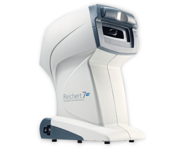
5. Eye pressure measurement
Intraocular pressure (IOP) is measured to screen for glaucoma, a condition that can lead to blindness if untreated. Normal IOP ranges from 10-21 mmHg, and higher values may indicate risk. We use a non-contact air puff test (Reichert 7 IOP) for a quick and accurate assessment, even for post-LASIK patients.
6. Additional tests
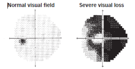
A) Visual Field Assessment
A visual field test may be recommended for individuals with:
- A family history of glaucoma
- Elevated IOP readings
This test detects dysfunction in central and peripheral vision, which may be caused by glaucoma, stroke, pituitary disease, brain tumors, or neurological conditions.
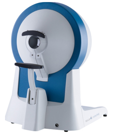
B) Axial Length Measurement
Axial length measurement is an essential tool for myopia management, particularly in children and young adults. It measures the length of the eyeball from the cornea to the retina.
Why is it important?
- The axial length of the eye is directly related to myopia progression. A longer axial length increases the risk of retinal detachment, myopic macular degeneration, and other sight-threatening conditions.
- Regular monitoring helps us assess the effectiveness of myopia control strategies, such as Ortho-K lenses, myopia control spectacles or contact lenses.
How is it measured?
- We use advanced optical biometers that accurately measure axial length in micrometers without physical contact, ensuring a comfortable and precise assessment.